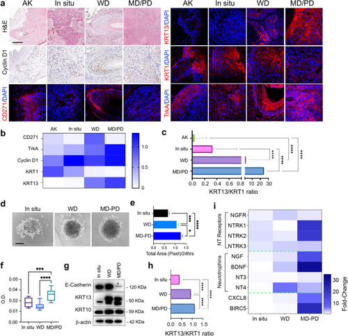
CD271 expression in cSCC biopsies and spheroids a Hematoxylin and Eosin (H&E) staining of AK, in situ, WD and MD/PD cSCC biopsies. CD271, Cyclin D1, TrkA, KRT1 and KRT13 expression evaluated by immunohistochemistry or immunofluorescence. Scale bar 100 µm. b Immunohistochemistry and immunofluorescence staining scored as follow: 0 (no cells; positivity to 20% of cells positivity), 0.1–0.75 (30–50% of cells positivity; 1–1.5 (60% to 100% of cells positivity). c Graphical representation of the KRT13/KRT1 intensity ratio. d Representative pictures of in situ, WD, and MD/PD spheroids. Scale bar = 100 μm. e Spheroid total area measured by ImageJ software and f spheroid viability evaluated by MTT assay. g Expression of E-cadherin, KRT13, and KRT10 evaluated in patient-derived spheroids by western blotting. β-actin was used as control. h Graphical representation of KRT13 and KRT10 relative protein expression ratio. i NTs and NTRs mRNA expression evaluated in patient-derived spheroids by qPCR. Heatmap created by Prism Graph pad software. β-actin was used as a housekeeping gene. For all experiments, the results are shown as mean ± SD of three independent experiments. Statistical analysis was performed using the two-way ANOVA. *0.01 < p < 0.05, ***0.0001 < p < 0.001, ****p < 0.0001
|
