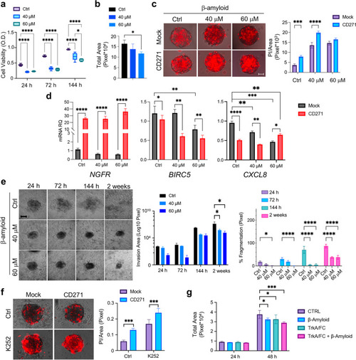
Effects of CD271 activation on cSCC spheroid viability and invasion a Cell viability (MTT assay) and b total area of SCC13 spheroids (n≥6) after β-Amyloid (40 μM and 60 μM) treatment as compared to control (PBS). c Left panel: Propidium Iodide (PI) staining (red)/brightfield on mock vs CD271-overexpressing SCC13 spheroids after β-Amyloid treatment as compared to control (PBS) at 48 h. Right: Bar chart representing PI-stained area (Pixel) (n = 3). Scale bar = 50 μm dNGFR, BIRC5 and CXCL8 mRNA levels (n = 8). e Left panel: Representative images of SCC13-derived implanted spheroid treated with β-Amyloid or PBS (control) and monitored for 2 weeks (scale bar = 50 μm). Middle panel: total invasion area. Right panel: percentage of fragmentation. f Left panel: PI staining/brightfield of CD271 and mock SCC13 spheroids treated with K252 (200 nM) or DMSO at 48 h. Right panel: Quantification of the PI staining/Total area ratio. Scale bar = 50 μm g Total area of SCC13 spheroids treated with TrkA/Fc (2 µg/ml), βAmyloid (40 μM) or the combinations TrkA/FC/βAmyloid (n = 3). Results are shown as mean ± SD of three independent experiments. Statistical analysis was performed using the two-way ANOVA and multiparametric T-test. *0.01 < p < 0.05, **0.001 < p < 0.01, ***0.0001 < p < 0.001, ****p < 0.0001
|
