|
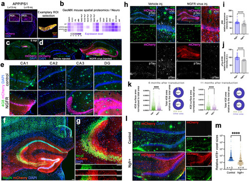
Ngfr reduces Aβ42 load and phosphorylated Tau in the hippocampus of APP/PS1dE9 mice. a Representative image from GeoMx spatial proteomics mouse brain section and region of interest (ROI) at the SGZ. b Heatmap showing the detection levels of selected proteins after Ngfr transduction in wild type and APP/PS1dE9 mice. c Representative APP/PS1dE9 mouse brain section immunostained for 4G8 and mCherry with DAPI counterstain at 6 months after transduction with Lv16 in one DG. d High-magnification image from c. e Comparison of amyloid load in CA1, CA2, CA3 and DG regions of control and Ngfr-transduced hemispheres. f Double immunostaining for NeuN and mCherry with DAPI counterstain at 6 months after transduction. g Higher magnification image of f. Individual channel panels indicate overlapping mCherry and NeuN. h Quantification of amyloid load in terms of normalized amyloid immunoreactivity by comparing control (v) versus Ngfr (Lv16) transduction. n = 5. i. 4G8, mCherry, phosphorylated Tau-S199 immunostaining with DAPI counterstain in control and Ngfr-injected DGs. Individual fluorescence channels and close-up images are also shown. j Quantification of pTau-S199 in terms of normalized fluorescence by comparing control (v) versus Ngfr (Lv16) transduction. n = 5. k 4G8 surface area quantification and relative 4G8 area graphs for 6 months and 11 months after Ngfr transduction. l Double immunolabeling for AT8 and mCherry with DAPI counterstain in control and Ngfr+ animals. Individual panels show single fluorescent channels. m Quantification of relative AT8 immunoreactivity. Ngfr reduces the prevalence of AT8. Error bars represent standard error of the means. Scale bars equal 100 μm.
|