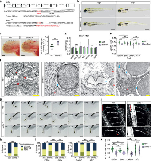
amfra-/- zebrafish larvae show alterations in lipid metabolism and endoplasmic reticulum morphology, abnormal touch-evoked escape response and motor neuron branching defects that can be rescued by statin treatment. a Schematic drawing of the amfra locus in zebrafish, the gRNA used and the generated mutation, causing a 5 bp frameshift in the first coding exon of amfra. Coding exons, black; non-coding exons, white. b Representative bright-field images of wild type and amfra-/- larvae at 3 dpf and 5 dpf. amfra-/- larvae appear morphologically similar to wild-type (WT) larvae at both developmental stages. Scale bars = 500 µm. c Representative images and quantification of ORO staining of 3 dpf larvae, dotted line indicating the region of interest (ROI) used for quantifications. Circles show individual values for each larva, n = 6 larvae per genotype (WT, green; amfra-/-, purple). Error bars represent SD (Kruskal–Wallis test, **p < 0.01). Scale bars = 100 µm. d qRT-PCR expression analysis for selected cholesterol metabolism genes, in brains of 5 dpf control and amfra-/- larvae (n = 10 brains per sample, 4 biological replicates for WT larvae and 5 biological replicates for amfra-/-, from 2 independent experiments, with each biological replicate measured in two technical replicates). Bar plot showing the mean fold change for the indicated genes compared to control larvae, normalized for the housekeeping gene eef1a1. WT, green. amfra-/-, purple. Error bars represent SD (Kruskal–Wallis test, ***p < 0.001). e Violin plot showing the length of wild type (WT) and amfra-/- larvae at 5 dpf. Larvae are either treated starting from 8 hpf onwards with simvastatin (SMV) or atorvastatin (ATV), or with their respective vehicle controls ethanol or DMSO. Whereas statin treatment does not influence the length of WT larvae, it significantly increases the length of amfra-/- larvae, partially rescuing the observed length deficit of amfra-/- larvae compared to WT. n > 20 per genotype and treatment group (Dunn’s Multiple Comparison test, **p < 0.01; ***p < 0.001). f Electron microscopy of brains from WT and amfra-/- larvae at 5 dpf. amfra-/- larvae show dilated rough endoplasmic reticulum decorated with ribosomes (blue *) and an expanded perinuclear space (blue #) as compared to controls (red * and #, respectively). Many cells in the amfra-/- samples have less densely stained cytoplasm (example indicated with a blue $). m = mitochondrion (normal appearing) and g = Golgi apparatus (normal appearing). Scale bars = 500 nm. g Representative bright-field images for the touch-evoked escape response (upper row: normal response; middle row: delayed response; bottom row: no response) of 3 dpf WT and amfra-/- larvae. Scale bars = 500 µm. h Quantification of the touch-evoked escape response in WT and amfra-/- larvae at 3 dpf, n > 45 per genotype, from 3 experimental replicates (Chi-square test, ***p < 0.001). i As H, but now for WT and amfra-/- larvae treated with simvastatin (SMV) or atorvastatin (ATV) and their respective vehicle controls, ETOH and DMSO (Chi-square test, **p < 0.01; ***p < 0.001; ns = not significant). j Immunostaining for acetylated tubulin in WT and amfra-/- embryos. Shown are max projections from z-stacks of 2 dpf embryos, acquired from lateral view of the middle of the trunk. The region indicated by the red rectangle is shown in enlargement in the insert below. amfra-/- embryos show reduced axon branching in ventral motor neurons in comparison to control (red arrow indicates a branching). Scale bars = 50 µm. k Violin plot showing the quantification of axon branching of ventral motor neurons in WT (green) and amfra-/- embryos (purple), treated with vehicle (ETOH or DMSO) or statin (SMV or ATV); n = 6 embryos per group; for each embryo, two axons were quantified. Black circle, median; black line, SD (Dunn’s Multiple Comparison test, **p < 0.01; ***p < 0.001; ns = not significant)
|

