|
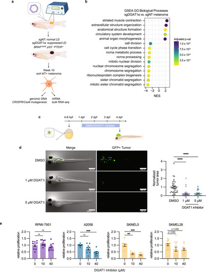
Inhibiting lipid droplet formation suppresses tumor growth and cell cycle progression. a Schematic of zebrafish TEAZ and sort for genomic DNA and mRNA. Transgenic casper;mitfa:BRAFV600E;p53−/− zebrafish were injected with tumor-initiating plasmids and electroporated to generate BRAFV600Ep53−/−PTENko melanomas with normal or suppressed lipid droplet formation. Melanoma cells were sorted to extract genomic DNA and mRNA. b Top 15 most significant (multi-level Monte Carlo by Benjamini-Hochberg adjusted p values) GO Biological Processes positively and negatively enriched in sgDGAT1a tumors. c Schematic of ZMEL-LD blastula transplant assay. d Representative images and tumor area quantification of blastula transplant. (mean ± SEM, DMSO n = 31 biologically independent replicates, 1 µM n = 27 biologically independent replicates, 5 µM n = 19 biologically independent replicates over n = 3 biologically independent experiments). Statistics via Kruskal Wallis with Dunn’s multiple comparison test. **** p < 0.0001. e Relative cell proliferation in RPMI-7951 (U/NC), A2058 (T), SKMEL5 (M), and SKMEL28 (M) treated with indicated DGAT1i inhibitor concentrations in nutrient limited media. (mean ± SEM, RPMI-7951 n = 9 biologically independent replicates, A2058 n = 9 biologically independent replicates, SKMEL5 n = 9 biologically independent replicates, SKMEL28 n = 12 biologically independent replicates over n = 3 biologically independent experiments). Statistics via Kruskal Wallis with Dunn’s multiple comparison test. ** p < 0.01, *** p < 0.001. Figure 6a, c was created with Biorender.com.
|