
FIGURE
Figure 2
- ID
- ZDB-FIG-230416-94
- Publication
- Tamburello et al., 2023 - Preclinical Evidence of Progesterone as a New Pharmacological Strategy in Human Adrenocortical Carcinoma Cell Lines
- Other Figures
- All Figure Page
- Back to All Figure Page
Figure 2
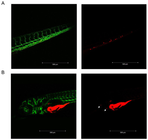
MUC-1 and TVBF-7 ACC cells induced metastases formation in zebrafish embryos. (A) Enlargement of the embryo tail with metastasized MUC-1 cells. (B) A representative acquisition of the metastases of TVBF-7 cells in the pericardial area of the embryo is shown. Cells are labeled with a red fluorescent lipophilic dye while the embryos’ endothelium is labeled with a green fluorescent protein reporter driven by the kdrl promoter. Images were acquired at 120 hpf using a Zeiss LSM 510 META confocal laser scanning microscope at 10× magnification. |
Expression Data
Expression Detail
Antibody Labeling
Phenotype Data
Phenotype Detail
Acknowledgments
This image is the copyrighted work of the attributed author or publisher, and
ZFIN has permission only to display this image to its users.
Additional permissions should be obtained from the applicable author or publisher of the image.
Full text @ Int. J. Mol. Sci.