
Figure 5
- ID
- ZDB-FIG-230416-5
- Publication
- Gopal et al., 2023 - Development of a Triple-Negative Breast Cancer Leptomeningeal Disease Model in Zebrafish
- Other Figures
- All Figure Page
- Back to All Figure Page
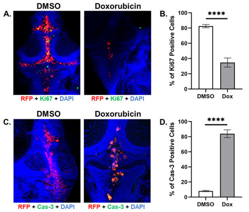
Doxorubicin treatment decreased cell proliferation and increased cell death compared to DMSO vehicle in zebrafish MDA-MB-231 TNBC xenografts. (A). Z-stack analysis combining red (MDA-MB-231-Luc/RFP cells), green (Ki67-stained cells), and blue (DAPI-nuclear-stained cells) filter images show that xenograft samples treated with DMSO vehicle exhibited more Ki67 staining than samples treated with doxorubicin at 8 dpi. (B). Doxorubicin treatment reduced the percentage of Ki67-positive cells compared to DMSO-vehicle-treated cells at 8 dpi. (C). Z-stack analysis combining red (MDA-MB-231-Luc/RFP cells), green (cleaved-caspase-3-stained cells), and blue (DAPI-nuclear-stained cells) filter images show that xenograft samples treated with DMSO vehicle exhibited more cleaved caspase-3 staining than samples treated with doxorubicin at 8 dpi. (D). Doxorubicin treatment increased the percentage of cleaved-caspase-3-positive cells compared to DMSO-vehicle-treated cells at 8 dpi. Key: RFP = red fluorescent protein; DAPI = 4′,6-diamidino-2-phenylindole; Cas-3 = caspase-3; Dox = doxorubicin. N = 10; “****” = p < 0.0001; Error bars = SEM. |