
Fig. 4
- ID
- ZDB-FIG-230410-4
- Publication
- Kourpa et al., 2022 - 15-keto-Prostaglandin E2 exhibits bioactive role by modulating glomerular cytoarchitecture through EP2/EP4 receptors
- Other Figures
- All Figure Page
- Back to All Figure Page
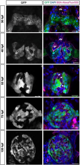
Fig. 4. Glomerular morphogenesis occurs in distinct steps. Zebrafish glomerular developmental stages. Representative high-resolution confocal microscopy images (single confocal section) of the Tg[wt1b:eGFP] developing zebrafish embryonic kidney at different developmental stages, 30 hpf (nephron primordia), 48 hpf, 52 hpf, 72 hpf and 120 hpf in an order from top to bottom. The podocytes and parietal epithelial cells in the glomeruli in grey (left), in green (in merge, right), endothelial cells of glomeruli capillaries in magenta (BSA-AlexaFluor555, in merge), cells nuclei in blue (DAPI, in merge). At 52 hpf, podocyte protrusions (arrowheads) start surrounding the capillaries to form the mature GFB. Dorsal aorta (da) and glomerular cleft (gc) are observed at the early developmental stages of the glomerulus. Scale bar = 10 μm. (For interpretation of the references to colour in this figure legend, the reader is referred to the web version of this article.) |