FIGURE
            Figure 5
- ID
- ZDB-FIG-230401-7
- Publication
- Pezzotta et al., 2023 - Combined Inhibition of Hedgehog and HDAC6: In Vitro and In Vivo Studies Reveal a New Role for Lysosomal Stress in Reducing Glioblastoma Cell Viability
- Other Figures
- All Figure Page
- Back to All Figure Page
                
                    
                        Figure 5


| 
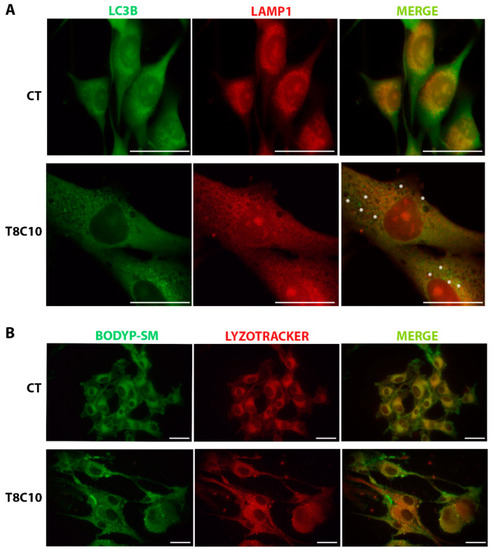
 LC3 and LAMP1 colocalization and BODIPY-SM distribution were impaired following Hh and HDAC6 inhibition. (A) Double immunostaining against LC3 and LAMP1 of U87-MG cells treated in the presence or absence of both 8 μM of TubA and 10 μM of cyclo. Asterisks show some LC3 puncta, indicating autophagosomes, that did not colocalize with the red ones, indicating lysosomes. (B) BODIPY-SM and LysoTracker Red DND-99 staining of U87-MG cells treated in the presence or absence of both 8 μM of TubA and 10 μM of cyclo. CT—control, TubA/T—tubastatin A; cyclo/C—cyclopamine. Scale bar indicates 40 μm. | 
                
                    
                        Expression Data


                        Expression Detail


                        Antibody Labeling


                        Phenotype Data


                        Phenotype Detail


                        Acknowledgments


This image is the copyrighted work of the attributed author or publisher, and
ZFIN has permission only to display this image to its users.
Additional permissions should be obtained from the applicable author or publisher of the image.
      Full text @ Int. J. Mol. Sci.


        