
Figure 4
- ID
- ZDB-FIG-230331-64
- Publication
- Yuan et al., 2023 - Roles of miR-196a and miR-196b in Zebrafish Motor Function
- Other Figures
- All Figure Page
- Back to All Figure Page
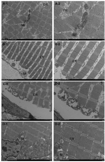
Transmission electron microscopy results of dorsal muscle tissue of miR-196a-1 and miR-196b knockout zebrafish. The zebrafish muscle tissue was damaged after miR-196a-1 or miR-196b gene knockout and mainly showed irregular morphology of muscle cells, irregular arrangement of myofibrils, sarcoplasmic reticulum dilatation, and mitochondrial swelling. (A): Wild-type zebrafish; (B): zebrafish with miR-196a-1 gene knockout; (C): miR-196b gene knockout zebrafish; (D): zebrafish with miR-196a-1 and miR-196b gene knockout; left panels: 2500×, right panels: 5000×. ER is sarcoplasmic reticulum, M is mitochondria, and N is muscle nucleus. |
| Fish: | |
|---|---|
| Observed In: | |
| Stage: | Adult |