
Fig. 3
- ID
- ZDB-FIG-230331-171
- Publication
- Xu et al., 2023 - Depletion of stearoyl-CoA desaturase (scd) leads to fatty liver disease and defective mating behavior in zebrafish
- Other Figures
- All Figure Page
- Back to All Figure Page
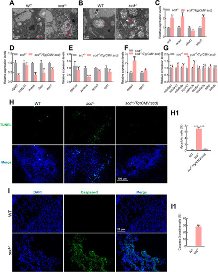
A: Scanning electron micrographs of WT and scd -/- liver sections showing increased lipid droplets in mutant livers. Scale bars: 10 μm. B: Scanning electron micrographs of WT and scd -/- liver sections showing shrunken mitochondria in mutant liver cells. Scale bars, 2 μm. C: Expression levels of scd, rxraa, elovl2, and elovl5 in scd -/- and scd -/- /Tg(CMV:scd) livers at 3 mpf (RT-qPCR). D: Expression levels of fatty acid synthesis-related genes dgat2, srebf1, acaca, fasn, and acc1 were significantly lower in scd -/- /Tg(CMV:scd) livers than in scd -/- livers at 3 mpf (RT-qPCR). E: Expression levels of oxidation-related genes pparαa, pparαb, acox3, and cpt1 in scd -/- and scd -/- /Tg(CMV:scd) livers at 3 mpf (RT-qPCR). F: Expression levels of transfer-related genes apoa1 and apob in scd -/- and scd -/- /Tg(CMV:scd) livers at 3 mpf (RT-qPCR). G: Expression levels of oxidative phosphorylation-related genes ndufab1, ndufa10, and ndufa9a in scd -/- and scd -/- /Tg(CMV:scd) livers at 3 mpf (RT-qPCR). H: Detection of liver cell apoptosis in WT, scd -/- , and scd -/- /Tg(CMV:scd) fish ( n=3, two sections for each sample) at 3 mpf with TUNEL staining, with DAPI blue indicating nucleus and TUNEL green indicating apoptosis signal. Scale bars: 100 μm. H1: Number of apoptotic cells (%) ( n=3, two sections for each sample). I: Immunofluorescence detection of WT and scd -/- liver sections ( n=3, two sections for each sample) with Caspase-3 antibody. Scale bars: 25 μm. I1: Number of Caspase-3-positive cells (%) ( n=3, two sections for each sample). All values are mean± SEM. Student t-tests were used. *: P<0.05; **: P<0.01; ***: P<0.001; ns: No significance. |