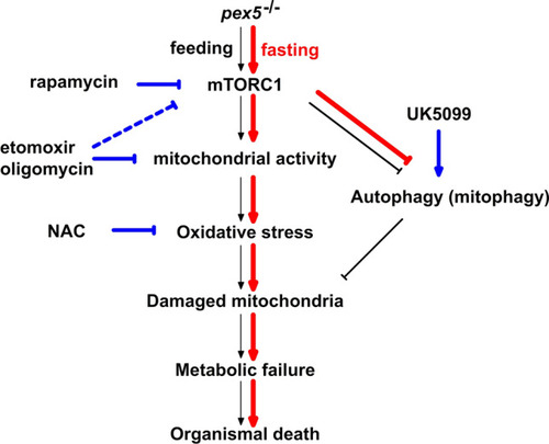
Fig. 8
- ID
- ZDB-FIG-230228-82
- Publication
- Bhandari et al., 2023 - Loss of pex5 sensitizes zebrafish to fasting due to deregulated mitochondria, mTOR, and autophagy
- Other Figures
- All Figure Page
- Back to All Figure Page
|
A model to illustrate the fasting-induced early mortality in pex5−/− in this study. Events occurring primarily in the pex5−/− liver under normal fed conditions are shown in black arrows. Briefly, due to dysfunctional peroxisomes, metabolic burden in pex5−/− induces compensatory activation of mitochondria which may then lead to mitochondrial damage and subsequent metabolic failure and organismal death. The entire process may run slow and take almost one month presumably due to the availability of nutrients under fed conditions. However, the process becomes highly facilitated under fasting conditions. mTORC1 becomes repressed in the fasted WT liver but not in the pex5−/− liver. In the fasted pex5−/− liver, mitochondrial activities are upregulated by unrepressed mTORC1 whose activity may also regulated by mitochondrial activity. Further, abnormally high mitochondrial activities may rapidly induce mitochondrial dysfunction and lead to accumulation of damaged mitochondria, which may not be replenished due to the limited autophagic flux. The undesirable condition upon fasting then leads to metabolic failure and results in early mortality of pex5−/−. The events strengthened upon fasting are shown by red arrows. The points of action of drugs that either activate or inhibit the process are shown in blue |

