
Fig. 2
- ID
- ZDB-FIG-230227-2
- Publication
- Bump et al., 2022 - Osteoblasts pattern endothelium and somatosensory axons during zebrafish caudal fin organogenesis
- Other Figures
- All Figure Page
- Back to All Figure Page
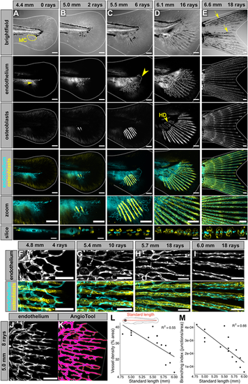
Stages of endothelial outgrowth, plexus formation, and remodeling during caudal fin development. (A-E) Lateral views of confocal projections of caudal fins fixed at the indicated stages expressing transgenic markers for osteoblasts [Tg(sp7:mCherry-NTR)] and endothelium [Tg(fli1a:EGFP)]. Dotted lines denote caudal fin margins. ‘Zoom’ panels show cropped, enlarged regions of merged images. ‘Slice’ panels represent reconstructed orthogonal views of the developing plexus and ray-associated osteoblasts. The yellow arrow in A indicates the ventral endothelial sprout. The yellow outline in A indicates the mesenchymal condensation (MC), visible under brightfield. The yellow arrowhead in C indicates the dorsal endothelial loop. The yellow arrowhead in D indicates the hypural diastema (HD), the future location between hypurals 2 and 3. The yellow arrows in E point to examples of melanophores tracking along bony rays. (F-I) Confocal projections of osteoblasts [Tg(sp7:mCherry-NTR)] and endothelium [Tg(fli1a:EGFP)], capturing endothelial remodeling around the osteoblasts at the indicated stages. Note that the initially disorganized, web-like plexus progressively remodels to its mature linear, ray-aligned morphology. (J,K) Example of an endothelial [Tg(fli1a:EGFP)] image (J), segmented and analyzed in AngioTool (K). Blue dots denote branchpoints. (L,M) Quantification of density and branching of the caudal fin endothelium relative to standard length. Scale bars: 100 μm (A-E); 20 μm (A-E, slice panels); 50 μm (F-J). |