
Fig. 1
- ID
- ZDB-FIG-230227-1
- Publication
- Bump et al., 2022 - Osteoblasts pattern endothelium and somatosensory axons during zebrafish caudal fin organogenesis
- Other Figures
- All Figure Page
- Back to All Figure Page
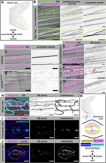
Somatosensory innervation of the adult caudal fin. (A) Schematic of the adult zebrafish caudal fin anatomy, composed of 18 bony rays. Distal (posterior) is to the right in this and subsequent lateral views. (B) Lateral view of adult caudal fin, fixed and immunostained for peripheral axons (zn-12 antibody) and acetylated tubulin. Insets show high-magnification images of a ray bifurcation. Note that zn-12 preferentially stains axon endings, whereas acetylated tubulin preferentially labels axon bundles. (C,D) Representative images of caudal fin bony rays (DIC) and acetylated tubulin staining in adult fins of the indicated genotypes. (E-G) Caudal fin bony rays (DIC) and somatosensory axons (SS axons) of an adult Tg(p2rx3a>mCherry) fish. Arrowheads in F illustrate an axon branching to exit the ray segment. Note the increased density of somatosensory axons at the dorsal-most fin edge in G as indicated by the brackets. (H) Single caudal fin bony ray (DIC), somatosensory axons (SS axons) and endothelium of an adult Tg(p2rx3a>mCherry);Tg(fli1a:EGFP) fish. Arrowheads denote a SS axon entering or exiting the bony ray, not associated with endothelial exit points (arrows mark one such example). (I) Transverse cross-section of a single bony ray of an adult Tg(p2rx3a>mCherry);Tg(fli1a:EGFP) fish. Note the lack of apparent association between axons and endothelium within the intra-ray space. (J) Transverse cross-section of a single bony ray of an adult Tg(p2rx3a>mCherry) fish, immunostained with the zns-5 antibody to label osteoblasts. Note the close juxtaposition of axons and intra-ray osteoblasts. (K) Schematic illustrating the anatomy of a transverse view of a single bony ray based on our results. Scale bars: 100 μm (B-G); 50 μm (H); 25 μm (I,J). |