
Figure 5
- ID
- ZDB-FIG-230211-152
- Publication
- Oderberg et al., 2023 - Biliary epithelial cells are facultative liver stem cells during liver regeneration in adult zebrafish
- Other Figures
- All Figure Page
- Back to All Figure Page
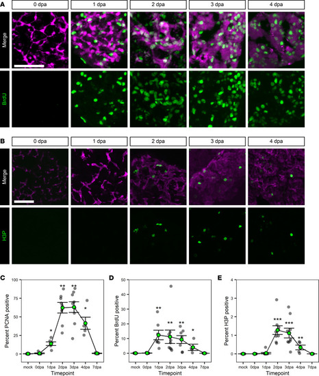
Proliferation of BECs during regeneration.
(A and B) Immunofluorescence in adult liver showing markers of biliary origin (magenta) and proliferation/cell cycling (green); mCherry (magenta) and BrdU (green) (A) or Anxa4 (magenta) and H3P (green) (B) signal for animals regenerating from hepatocyte ablation. Time points shown are BrdU: 0 dpa (n = 7), 1 dpa (n = 7), 2 dpa (n = 9), 3 dpa (n = 7), and 4 dpa (n = 4); H3P: 0 dpa (n = 9), 1 dpa (n = 9), 2 dpa (n = 9), 3 dpa (n = 9), and 4 dpa (n = 5). There is a burst in proliferation ranging from 1 to 4 dpa. Scale bars: 50 μm. (C–E) Line graph of the percentages of nuclei positive for PCNA (C), BrdU (D), or H3P (E) over time. Gray dots mark the average value for an animal; green-filled dot marks the average of the animal values. Data are shown as mean ± SEM. For PCNA and BrdU, the earliest significant change as compared with mock sample occurs at 1 dpa. For H3P, the earliest significant change is at 2 dpa. Significance was determined using the Wilcoxon Rank Sum test, and P values were adjusted for multiple hypothesis testing using a Bonferroni correction. *P < 0.05, **P < 0.01, ***P < 0.001. |