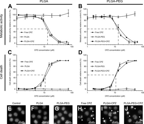
Fig. 7
|
CPZ-loaded PLGA nanoparticles have equal cytotoxicity towards AML cells compared to free CPZ. MOLM-13 cells were incubated for 24 h with various concentrations of either free CPZ, empty or CPZ-loaded nanoparticles. (A,C) are data with PLGA nanoparticles, and (B,D) are with PLGA-PEG nanoparticles. The cells were incubated with the different additions for 24 h before their viability relative to control was measured by the metabolic conversion of the WST-1 reagent. Following the plate reading for WST-1, the cells were fixed in 2% buffered formaldehyde (in pH 7.4 PBS containing Hoechst 33342). Using fluorescence microscopy, cells were counted for apoptosis and adjusted for control. The data in A-D are the averages and standard deviations from triplicate experiments. (E-J): Fluorescence microscopy images of Hoechst-stained nuclei of cells after 24 h of the different treatments. Arrows indicate typical apoptotic cells. Scale bars represent 20 µm. |

