
Fig. 5
- ID
- ZDB-FIG-221223-13
- Publication
- Qin et al., 2021 - Uptake of oxidative stress-mediated extracellular vesicles by vascular endothelial cells under low magnitude shear stress
- Other Figures
- All Figure Page
- Back to All Figure Page
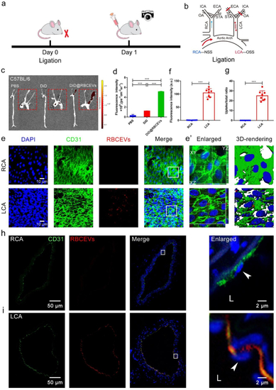
OSS promotes the uptake of RBCEVs by endothelial cells in C57BL/6 mice. (a) Schematic of ligation establishment. (b) LCA of mice was ligated and immediately injected with DiD@RBCEVs. (c) Small animal optical imaging system. (d) Quantitative analysis of (c). (e) En face immunofluorescence images. OSS increased the uptake of RBCEVs by endothelial cells. (f) Quantitative analysis of mean fluorescence intensity and (g) uptake ratio. RCA: non-ligation; LCA: ligation for 1 day, injected once. The mice in each group were sacrificed 1 h after the last injection. (e’) Imaris 3D rendering. (n = 8) (scale bar = 10 μm). (h–i) Immunofluorescence images and transection of the carotid artery. The white box indicates that the picture is locally enlarged, as indicated by the white arrow. LCA: ligation for 1 day, injection for 1 time, RCA: unligation. The mice in each group were sacrificed 1 h after the last injection. L: lumen. (n = 8) (scale bar = 50 μm). Significance is indicated as P < 0.001 (***). |