
Figure 1.
- ID
- ZDB-FIG-221211-309
- Publication
- Nelson et al., 2022 - The developmental progression of eight opsin spectral signals recorded from the zebrafish retinal cone layer is altered by the timing and cell type expression of thyroxin receptor β2 (trβ2) gain-of-function transgenes
- Other Figures
- All Figure Page
- Back to All Figure Page
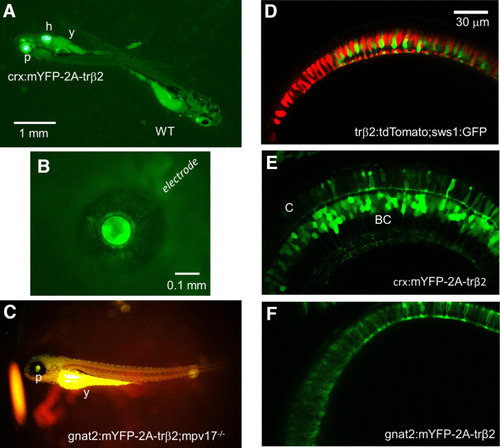
Reporter fluorescence of zebrafish larvae. A, Larvae from outcrosses of crx:mYFP-2A-trβ2 heterozygotes are either heterozygous or wild-type controls (WT). The heterozygotes are recognized by both pupil (p) and heart (h) fluorescence. The yolk sac (y) is autofluorescent. B, The cornea of a 5-d transgenic eye isolated from a crx:mYFP-2A-trβ2 larva is penetrated with a patch electrode for ERG recordings. The eye is <0.5-mm diameter. C, The gnat2:mYFP-2A-trβ2;mpv17−/− gain-of-function phenotype is studied on a roy orbison (mpv17−/−) background strain. The darkly pigmented, nonreflective iris of this control strain aids in visualizing the dim transgenic fluorescence of the pupil (p). The yoke (y) is autofluorescent. D–F, Live confocal imaging of retinas in 6-dpf larvae. D, WT red (red) and UV (green) cone morphology visualized with trβ2:tdTomato and sws1:GFP fluorescent reporter transgenes. E, The mYFP construct in the crx:mYFP-2A-trβ2 transgene causes cones (C) and bipolar cells (BC) to fluoresce. F, The mYFP construct in the transgene gnat2:mYFP-2A-trβ2 marks only cone cells. |