
Fig. 7
- ID
- ZDB-FIG-221211-305
- Publication
- Morales-Curiel et al., 2022 - Volumetric imaging of fast cellular dynamics with deep learning enhanced bioluminescence microscopy
- Other Figures
- All Figure Page
- Back to All Figure Page
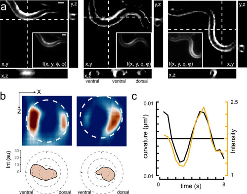
3D calcium imaging of freely moving animals.
a Sequence of reconstructed 3D images of a moving animal showing high-calcium activity at its contracted side. Images show a single plane of the reconstructed z-stack. Inset corresponds to the raw lightfield image. Scale bars = 40 μm. b Sideview image of the curvature-dependent calcium signal in muscles during ventral and dorsal body bends with warmer colors representing higher calcium signals. Scale bar = 20 μm. The polar plot shows the intensity distribution on the ventral and dorsal side. Dotted line corresponds to the circumference of the animal. c Intensity of the bioluminescent calcium indicator and curvature variation on the ventral side during animal crawling under the lightfield microscope. Black = curvature, yellow = calcium signal. |