
Fig. 3
- ID
- ZDB-FIG-221211-246
- Publication
- Wei et al., 2022 - Effects of bepridil on early cardiac development of zebrafish
- Other Figures
- All Figure Page
- Back to All Figure Page
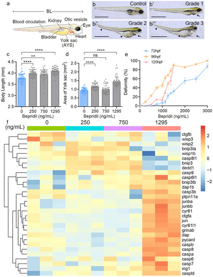
Morphological effects of zebrafish larvae after bepridil treatment. (a) Diagram of zebrafish larva and the internal organs. BL, body length. AYS, area of the yolk sac. (b-b’’’) Bepridil treatment caused different degrees of deformity at 120 hpf. Grade 1, grade 2 and grade 3 represent the levels of deformity from low to high. Black arrows showed the pericardial enlargement. Black arrowheads indicated swollen yolk sacs and curved tails. Scale bars = 1 mm. (c) Body lengths of zebrafish larvae at 120 hpf in each group. N = 45. (d) Quantification of AYS in zebrafish larvae at 120 hpf in control and different bepridil groups. N = 45. (e) Correlationship and zebrafish deformity of different bepridil concentrations at 72, 96, and 120 hpf. N = 45. (f) Heatmap of cell apoptosis-related genes expression pattern in different bepridil treatment groups |