
Fig. 8
- ID
- ZDB-FIG-221210-8
- Publication
- Walker et al., 2021 - Agrin/Lrp4 signal constrains MuSK-dependent neuromuscular synapse development in appendicular muscle
- Other Figures
- All Figure Page
- Back to All Figure Page
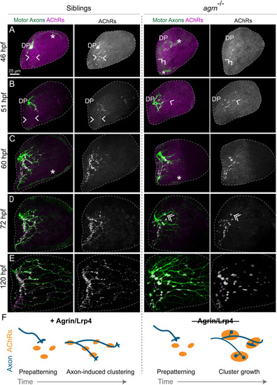
Agrin restricts presynaptic terminal and neural AChR cluster size. (A-E) Developmental time course from Tg(mnx1:GFP) larvae with motoneurons stained with α-Btx to label AChRs. (A,B) At 46 and 51 hpf, axons growing from the dorsal plexus (DP) have not yet innervated all prepatterned AChR clusters (single arrowheads). (C-E) Whereas small clusters are added as axons grow into the pectoral fin in sibling animals, clusters mainly increase in size in agrn mutants. Double arrow in D indicates presynaptic swelling colocalized with an AChR cluster. Only abductor innervation is shown, with the fin area outlined by the white-dashed line. Asterisks indicate endothelial or endoskeletal cells labeled in the green channel. (F) Schematic summarizing the developmental time course. Both siblings and agrn mutants look similar during the prepatterning stage. Incoming axons induce small AChR clusters in sibling animals, whereas, in agrn mutants, AChR clusters and axonal swellings increase in size over time. n=4-10 animals per genotype per time point. |