
Fig. 1
- ID
- ZDB-FIG-221210-1
- Publication
- Walker et al., 2021 - Agrin/Lrp4 signal constrains MuSK-dependent neuromuscular synapse development in appendicular muscle
- Other Figures
- All Figure Page
- Back to All Figure Page
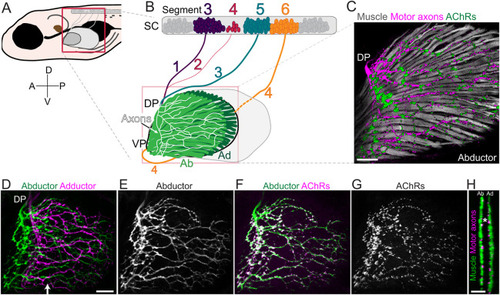
Pectoral fin anatomy. (A) Schematic of a 120 hpf (5 dpf) zebrafish larva. The boxed area indicates the region shown in more detail in B. (B) Schematic of pectoral fin motoneuron innervation. Motoneuron cell bodies are in spinal cord (SC) segments 3-6. Nerves 1-3 enter the fin at the dorsal plexus (DP), whereas nerve 4 (orange line) enters the fin at the ventral plexus (VP). All nerves innervate both the abductor (Ab) and adductor (Ad) muscles. (C) Ab innervation of a 120 hpf Tg(α-actin:GFP);Tg(Xla.Tubb:DsRed) pectoral fin stained with α-Btx to visualize muscle fibers, axons and AChRs (n=7 larvae). (D) Maximum projection of Tg(mnx1:GFP) innervation in the pectoral fin. Ab (green) and Ad (magenta) innervation patterns are pseudo-colored. (E) Ab innervation alone. (F) Ab innervation showing AChRs. (G) AChR labeling alone. n>77 WT pectoral fin images for D-G. (H) Cross-section of pectoral fin at approximate region marked by arrow in D. Asterisk marks the endoskeletal disk that separates the two muscles. A, anterior; D, dorsal; P, posterior; V, ventral. Scale bars: 25 μm. |