
Fig. 2
- ID
- ZDB-FIG-221128-41
- Publication
- Guan et al., 2021 - A specialized spinal circuit for command amplification and directionality during escape behavior
- Other Figures
- All Figure Page
- Back to All Figure Page
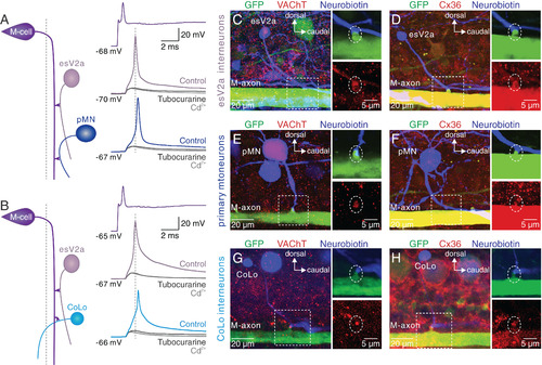
esV2a interneurons are recruited prior to pMNs and CoLo interneurons. (A) Triple whole-cell patch-clamp recording from an M-cell, an esV2a interneuron and a pMN. M-cell activation recruited first the esV2a interneuron and then the pMN. d-tubocurarine suppressed the firing of both neurons and only a Cd2+-insensitive eEPSP remained. (B) Triple whole-cell patch-clamp recording from an M-cell, an esV2a interneuron and a CoLo interneuron. The latter was recruited later than the esV2a interneuron. The cholinergic cEPSPs in both neurons were blocked by d-tubocurarine, leaving only an eEPSP remained that was unaffected by Cd2+. (C) Direct contact between the M-axon (green) and the axon of an esV2a interneuron (blue). VAChT (red) was expressed at the site of presumed synaptic contact (dashed box, Left). The large soma corresponds to a pMN expressing GFP in this line. (D) Expression of Cx36 (red) at the site of synaptic connections between the M-axon and esV2a interneuron (blue). (E) The expression of VAChT (red) was found at the presumed synaptic contact site between an M-axon (green) and a pMN (blue). (F) Cx36 (red) is expressed at the connection site between the M-axon and pMN. (G) The expression of VAChT (red) was found at the presumed synaptic contact site between an M-axon (green) and a CoLo interneuron (blue). (H) Expression of Cx36 (red) at the connection site between the M-axon and CoLo interneuron. The Right Upper and Lower panels in C–H represent magnification of the regions indicated by the dashed boxes in the Left panels. The white dotted circle indicates the synapses formed by the M-cell and spinal neurons. |