
Fig. 4
- ID
- ZDB-FIG-221127-45
- Publication
- Cudia et al., 2021 - NMR and EPR-DEER Structure of a Dimeric Guanylate Cyclase Activator Protein-5 from Zebrafish Photoreceptors
- Other Figures
- All Figure Page
- Back to All Figure Page
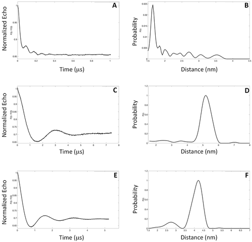
EPR-DEER intermolecular distances for the GCAP5 dimer. Representative EPR-DEER data of (A) GCAP5CL(A28C), (C) GCAP5CL(N56C), and (E) GCAP5CL(A69C) and corresponding distance distributions of (B) GCAP5CL(A28C), (D) GCAP5cl(N56C), and (F) GCAP5CL(A69C). GCAP5 samples were in the Ca2+-free, Mg2+-bound state. Similar DEER data were observed for GCAP5 in the Ca2+-bound state (not shown). A nitroxide spin-label (MTSSL) was covalently attached to the sole Cys residue in each mutant. The distance distributions and average intermolecular distances were calculated on the basis of the DEER data as described in Materials and Methods. The DEER intermolecular distances were measured to be 16 ± 1 Å [GCAP5CL(A28C) in panel B], 52 ± 3 Å [GCAP5CL(N56C) in panel D], and 41 ± 3 Å [GCAP5CL(A69C) in panel F]. |