
Fig. 6
- ID
- ZDB-FIG-221109-35
- Publication
- Zhou et al., 2022 - Dusp6 deficiency attenuates neutrophil-mediated cardiac damage in the acute inflammatory phase of myocardial infarction
- Other Figures
- All Figure Page
- Back to All Figure Page
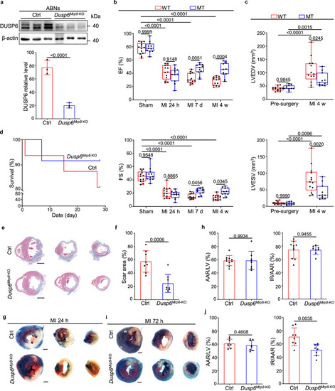
a Western blot and corresponding quantitative analysis of Dusp6 in ABNs from control and Dusp6Mrp8-KO mice. Control mice were Mrp8-Cre, and Dusp6Mrp8-KO mice were Mrp8-Cre, Dusp6f/f. β-actin served as internal control (n = 6 biological independent samples/group). Both blots were performed in parallel with the same samples. b Echocardiographic measurements of EF and FS for control (n = 13) and Dusp6Mrp8-KO (n = 11) mice before surgery, as well as 24 h, 7 days and 4 weeks after MI. c Echocardiographic measurements of LVEDV and LVESV for control (n = 13) and Dusp6Mrp8-KO (n = 11) mice before surgery and 4 weeks after MI. The box blots show center lines as median, box boundaries as upper and lower quartiles, and whiskers as minimum and maximum values. d Kaplan–Meier survival curves of control (n = 16) and Dusp6Mrp8-KO mice (n = 12) at 4w after MI. Representative Masson staining of heart sections (e) and quantitative analysis (f) of the fibrotic area from control and Dusp6Mrp8-KO hearts at 4 weeks after MI (n = 8/group. Scale bar, 1 mm). Representative TTC-Evans blue staining (g and i) and measurements of infarct size in control and Dusp6Mrp8-KO hearts at 24 h (h) (control: n = 10; Dusp6Mrp8-KO: n = 8) and 72 h (j) (n = 8/group) after MI. Scale bar, 1 mm. Quantitative data are presented as min to max with all points mean in b and c, and as values ± SD in a, f, h and j. One-way ANOVA with Tukey’s multiple comparison test (for b and c) and Two-sided unpaired T-test (for a, f, h and j) were used to calculate the presented p-values. Source data of a–d, f, h and j are provided in a Source Data File. Ctrl control, EF ejection fraction, FS fractional shortening, LVEDV left ventricular end-diastolic volume, LVESV left ventricular end-systolic volume, TTC triphenyl tetrazolium chloride, IR infarcted region, AAR area at risk, LV left ventricle. |