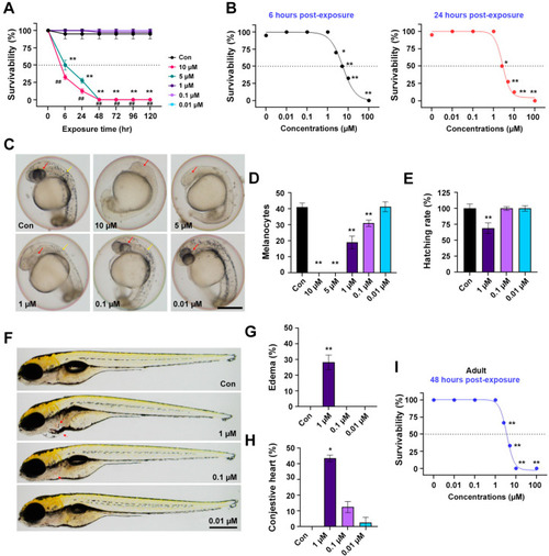
In vivo toxicity assay of 3f in the embryo and adult zebrafish. (A) Time-dependent survival rates throughout 0–120 h post exposure (hpe) in zebrafish embryos (n = 25 embryos/group). * p < 0.05, ** p < 0.01 compared to control and ## p < 0.01 compared to 5 hpe groups. (B) Concentration-dependent survival curves of 3f after continuous exposure for 6 h (left panel, black) and 24 h (right panel, red) in zebrafish embryos (n = 25 embryos/group). * p < 0.05 and ** p < 0.01. (C) Representative images of melanocyte development in zebrafish embryos exposed to 3f at 24 hpe. Scale bar, 0.25 mm. (D) The number of melanocytes in the head and trunk of zebrafish embryos was counted in three experiments. ** p < 0.01. (E) Hatching rate of zebrafish embryos after 48 h exposure to 3f (n = 25 embryos/group) was calculated. ** p < 0.01. (F) Malformation of zebrafish larvae exposed to 3f at 120 hpe was estimated. 3f-exposed zebrafish (1 µM) showed pericardial edema and congestive heart (red arrow). Scale bar, 0.5 mm. Quantification of pericardial edema (G) and congestive heart (H) in zebrafish larvae exposed to 3f for 120 h (n = 25 embryos/group) were shown. * p < 0.05 and ** p < 0.01. (I) Concentration-dependent survival curves of 3f after 3 consecutive days of exposure in adult zebrafish (n = 25 embryos/group) were displayed. Error bars indicated the standard deviation of the mean. ** p < 0.01.
|
