|
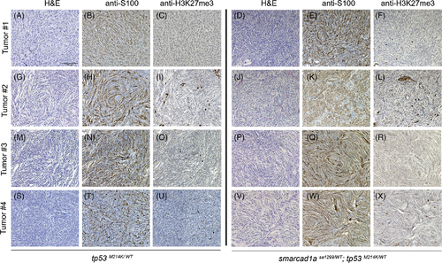
Confirmation of zebrafish MPNSTs with S100 and H3K27me3. Four random tumors from tp53 heterozygotes and double heterozygotes (tp53 and sa1299) were stained with S100 and H3K27me3 antibodies. (A–C, G–I, M–O, S–U): tp53 heterozygotes. (D–F, J–L, P–R, V–X): double heterozygotes. HE (hematoxylin and eosin) staining: A, D, G, J, M, P, S, V. IHC with S100 antibody: B, E, H, K, N, Q, T, W. IHC with H3K27me antibody staining: C, F, I, L, O, R, U, X. Some stained cells are non-Schwann cells in these sections, but they serve as a positive control for the antibody specificity. All the images were taken at the same magnification. Scale bar = 100 μm in panel A
|