
Fig. 5
- ID
- ZDB-FIG-221014-5
- Publication
- Yu et al., 2021 - Mechanism of gating and partial agonist action in the glycine receptor
- Other Figures
- All Figure Page
- Back to All Figure Page
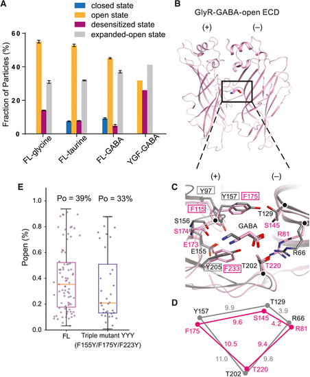
Particle distributions, binding pocket comparisons between GlyR and the α1β1γ2 GABA receptor-GABA complex and single channel open probabilities.
A. Fractions of GlyR particles in different states in the FL and YGF mutant, calculated by RELION and cisTEM. Error bars represent SEM. B. Extracellular domain (ECD) of the open state of the GABA complex in SMA. C. Superposition of the GABAAR (in grey; PDB code: 6DW1) and YGF mutant (in pink) structures highlights differences in the binding pockets. The Cα atoms of key residues are represented as spheres. D. Schematic diagram illustrating the distances of the Cα atoms of R81, F175, S145 and T220 in the GlyR and corresponding residues in the GABAAR structure. E. The open probabilities (Po) of the FL and the triple mutant (F115Y+F175Y+F223Y) elicited by 100 mM GABA. Error bars represent SEM and n≥6 cells for all experiments. |
Reprinted from Cell, 184, Yu, J., Zhu, H., Lape, R., Greiner, T., Du, J., Lü, W., Sivilotti, L., Gouaux, E., Mechanism of gating and partial agonist action in the glycine receptor, 957-968.e21, Copyright (2021) with permission from Elsevier. Full text @ Cell