
Fig. 3
- ID
- ZDB-FIG-220921-3
- Publication
- Chu et al., 2022 - In vivo drug discovery for increasing incretin-expressing cells identifies DYRK inhibitors that reinforce the enteroendocrine system
- Other Figures
- All Figure Page
- Back to All Figure Page
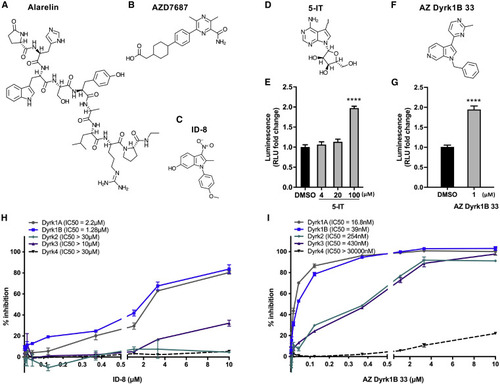
Figure 3. Structures of the hits and specificity of ID-8 and AZ Dyrk1B 33 on DYRKs (A–C) Chemical structures of the hits. Alarelin (A), AZD7687 (B), and ID-8 (C). (D–G) Chemical structures of the additional DYRK inhibitors followed up on, i.e., 5-IT (D) and AZ Dyrk1B 33 (F); and luminescence in Tg(gip:Nluc) zebrafish larvae treated from 3 to 6 days post fertilization (dpf) with 4, 20, and 100 μM 5-IT (E) or 1 μM AZ Dyrk1B 33 (G) (≥4 μM AZ Dyrk1B 33 was toxic to the zebrafish), normalized to DMSO. ∗∗∗∗p < 0.0001. (H and I) Specificity analyses of ID-8 (H) and AZ Dyrk1B 33 (I) on the DYRK-family of kinases by dose-response assessment in vitro. The SelectScreen biochemical kinase profiling assays were performed by ThermoFisher Scientific, using LanthaScreen for DYRK2 and Z′-LYTE for DYRK1A, DYRK1B, DYRK3, and DYRK4. See also Figure S2 and S3. |