
Fig. 6
- ID
- ZDB-FIG-220916-11
- Publication
- Mendieta-Serrano et al., 2022 - Slow muscles guide fast myocyte fusion to ensure robust myotome formation despite the high spatiotemporal stochasticity of fusion events
- Other Figures
- All Figure Page
- Back to All Figure Page
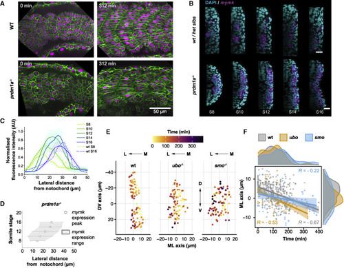
Figure 6. Loss of slow fibers results in the alteration of spatiotemporal dynamics of fast myocyte fusion (A) ubo mutant at 22-somite stage expressing Lyn-GFP (cell membrane, green) and H2B-mCherry (nuclei, magenta) with sibling control. Scale bars, 50 μm. Time t = 0 represents segment generation from the PSM. (B) Localization of mymk expression (magenta) visualized by FISH in a ubo mutant embryo at 22-somite stage co-stained with DAPI (nuclei, cyan), with sibling control. Scale bars, 20 μm. (C) Expression profile of mymk along the ML axis in different somite stages of ubo mutant embryos. Data from three embryos, with intensity normalized as Figure 5B. Dashed lines show comparison for wild-type embryos (from Figure 5B) in S8 and S16. Position 0 μm corresponds to the notochord/myotome boundary. (D) mymk expression domain (gray region, with mean ± SD shown by hexagons and error bars, respectively) in ubo mutants in the ML axis at different somite stages. Data from 3 embryos, distance as in (C). (E) Spatial distribution of fusion events in DV-ML axes in wild-type, ubo, and smo mutant embryos. Nuclei positions at the time of fusion color coded by time normalized to the first fusion event per individual myotome. Position 0 μm in each axis corresponds to segment center, determined by average cell position. (F) Timing of fusion events along ML axis in wild-type, ubo, and smo embryos. Darker colored lines represent linear fit, with lighter colored regions denoting the 95% CI. Time t = 0 min defined by the first fusion event, and ML position 0 μm corresponds to average cell position in ML axis for each segment. (E and F) Wild-type (179 fusion events from 9 myotomes from 3 embryos), ubo− (45 fusion events from 3 myotomes from 1 embryo), and smo (46 fusion events from 3 myotomes from 1 embryo). |
Reprinted from Developmental Cell, 57(17), Mendieta-Serrano, M.A., Dhar, S., Ng, B.H., Narayanan, R., Lee, J.J.Y., Ong, H.T., Toh, P.J.Y., Röllin, A., Roy, S., Saunders, T.E., Slow muscles guide fast myocyte fusion to ensure robust myotome formation despite the high spatiotemporal stochasticity of fusion events, 2095-2110.e5, Copyright (2022) with permission from Elsevier. Full text @ Dev. Cell