
Figure 2
- ID
- ZDB-FIG-220912-39
- Publication
- Routledge et al., 2022 - The scaffolding protein Flot2 promotes cytoneme-based transport of Wnt3 in gastric cancer
- Other Figures
- All Figure Page
- Back to All Figure Page
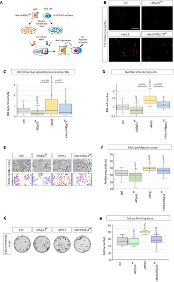
(A) Experimental protocol for measuring paracrine Wnt signalling activation. HFE cells expressing the SuperTOPFlash (STF) reporter, 7×TCF-NLS-mCherry, were cocultivated with AGS cells expressing indicated constructs. Fluorescence of STF mCherry reporter was measured after 48 hr and compared to untransfected control cells. (B) Representative images of STF reporter fluorescence for indicated conditions. Scale bar 100 µm. (C) Quantification of STF mCherry reporter fluorescence in HFE cells co-cultured with AGS (n per condition = 322, 394, 258, and 275). (D) Relative number of HFE cells per image after co-culture with AGS cells expressing indicated constructs. Significance calculated by Student’s t-test with Bonferroni correction for multiple comparisons. (n per condition = 28, 26, 27, and 17; n=number of images). (E) Representative images of proliferating, BrdU-stained (red); co-cultured AGS and HFE-145 cells, as described in (a). Cells were counterstained with haematoxylin (blue dots). Scale bar 100 µm. Complementary images show BrdU+ cells with red dots; blue dots mark BrdU- cells. (F) Quantification of BrdU-stained cells as a percentage of the population. Significance calculated by Student’s t-test with Bonferroni correction for multiple comparisons. (n per condition = 20, 20, 20, and 20; n=number of images). (G), Colony-forming assay of AGS cells. AGS cells were transfected with the indicated constructs and co-cultured with AGS-RFP cells for 2 days. After sorting, AGS-RFP expressing cells were plated at clonal density for 10–12 days; (H), Quantification of spherical colonies. Significance is calculated by Student’s t-test (n=9).
|