FIGURE

FIGURE 3

ID
ZDB-FIG-220903-3
Publication
Risato et al., 2022 - Hyperactivation of Wnt/β-catenin and Jak/Stat3 pathways in human and zebrafish foetal growth restriction models: Implications for pharmacological rescue
Other Figures
All Figure Page
Back to All Figure Page
FIGURE 3

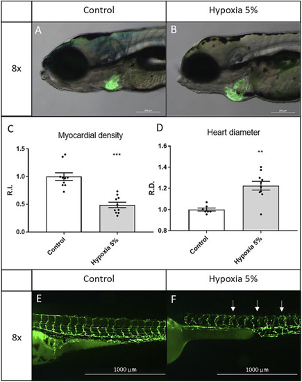
Long-term hypoxia induces cardiovascular modifications in zebrafish embryos/larvae. (A–D) After a 5-days incubation in 5% hypoxia, 6-dpf zebrafish larvae display enlarged heart and inflated myocardial layer, as evidenced by the fluorescence of the myocardial transgene Tg(tg:EGFP-myl7:EGFP)ia300. (E,F) After a 2-days incubation in 5% hypoxia, 3-dpf zebrafish embryos display disorganized intersegmental vessels (arrows), identified by the endothelial transgene Tg(fli1:EGFP)y1. 6-dpf larvae and 3-dpf embryos are in lateral view, anterior to the left. Sample size: n = 10; ** = p < 0.01; *** = p < 0.001; R.I. = Relative Intensity; R.D. = Relative Diameter. Test: Unpaired t-test.

Expression Data

Expression Detail
Antibody Labeling
Phenotype Data

Phenotype Detail
Acknowledgments
This image is the copyrighted work of the attributed author or publisher, and ZFIN has permission only to display this image to its users. Additional permissions should be obtained from the applicable author or publisher of the image. Full text @ Front Cell Dev Biol