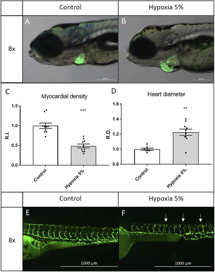
Long-term hypoxia induces cardiovascular modifications in zebrafish embryos/larvae. (A–D) After a 5-days incubation in 5% hypoxia, 6-dpf zebrafish larvae display enlarged heart and inflated myocardial layer, as evidenced by the fluorescence of the myocardial transgene Tg(tg:EGFP-myl7:EGFP)ia300. (E,F) After a 2-days incubation in 5% hypoxia, 3-dpf zebrafish embryos display disorganized intersegmental vessels (arrows), identified by the endothelial transgene Tg(fli1:EGFP)y1. 6-dpf larvae and 3-dpf embryos are in lateral view, anterior to the left. Sample size: n = 10; ** = p < 0.01; *** = p < 0.001; R.I. = Relative Intensity; R.D. = Relative Diameter. Test: Unpaired t-test.
Acknowledgments
This image is the copyrighted work of the attributed author or publisher, and
ZFIN has permission only to display this image to its users.
Additional permissions should be obtained from the applicable author or publisher of the image.
Full text @ Front Cell Dev Biol
Your Input Welcome
Thank you for submitting comments. Your input has been emailed to ZFIN curators who may contact you if
additional information is required.
Oops. Something went wrong. Please try again later.