
Fig. 8
- ID
- ZDB-FIG-220829-185
- Publication
- Romagnoli et al., 2022 - Design, Synthesis and Biological Investigation of 2-Anilino Triazolopyrimidines as Tubulin Polymerization Inhibitors with Anticancer Activities
- Other Figures
- All Figure Page
- Back to All Figure Page
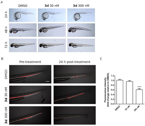
Effects of 3d treatment on the zebrafish xenotransplantation model (A). Compound 3d did not induce abnormal phenotypes or developmental anomalies in zebrafish embryos after 24, 48 and 72 h of incubation. DMSO-treated embryos were used as control. (B) Representative images of Tg(fli1:EGFP) zebrafish embryos (blood vessels shown in white) transplanted with DiI + HeLa cells (red). The embryos were treated with the indicated concentrations of 3d for 24 h and then the fluorescence intensity was quantified as depicted in panel (C). Data are expressed as mean ± SD (**** p < 0.001). Scale bar, 200 μm. Effects of 3d treatment on zebrafish embryos. No abnormal phenotypes or developmental defects were seen in comparison to DMSO-treated embryos (as a normal control) after 24, 48 and 72 h. (B) Effects of 3d treatment on the zebrafish xenotransplantation model. Representative images of Tg(fli1:EGFP) zebrafish embryos (blood vessels shown in white) transplanted with DiI + HeLa cells (red). Embryos were treated for 24 h with DMSO (control group), 30 nM 3d or 300 nM 3d. (C) Histograms represent the fluorescence intensity of the tumor xenografts, indicating total HeLa cells present in each embryo after a 24 h treatment with 3d at the indicated concentrations. Data are expressed as mean ± SD (**** p < 0.001). Scale bar, 200 μm. |