
FIGURE
Fig. 6
- ID
- ZDB-FIG-220810-22
- Publication
- Chen et al., 2022 - Leukocyte invasion of the brain after peripheral trauma in zebrafish (Danio rerio)
- Other Figures
- All Figure Page
- Back to All Figure Page
Fig. 6
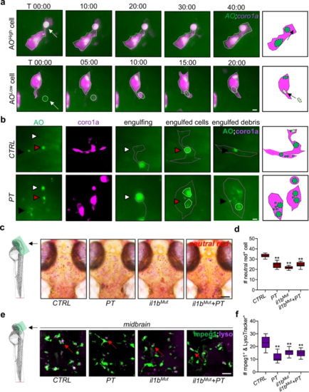
a, b Time-lapse light-sheet imaging showed the phagocytosis of AOhigh and AOlow cells (white arrow or arrowhead) by coro1a + leukocytes in the zebrafish brain at 1 dpt. c, d Neutral red staining showed microglial lysosome impairment in zebrafish brains with or without il1b mutation at 1 dpt. Two-way ANOVA with Tukey’s HSD post hoc test, **, p < 0.01 compared to CTRL. Scale bar, 40 µm. e, f LysoTracker staining showed mpeg1 + microglial lysosome impairment in the zebrafish brain at 1 dpt or under il1b mutation. Two-way ANOVA with Tukey’s HSD post hoc test, **, p < 0.01 compared to CTRL. Scale bar, 20 µm. |
Expression Data
Expression Detail
Antibody Labeling
Phenotype Data
| Fish: | |
|---|---|
| Condition: | |
| Observed In: | |
| Stage: | Day 4 |
Phenotype Detail
Acknowledgments
This image is the copyrighted work of the attributed author or publisher, and
ZFIN has permission only to display this image to its users.
Additional permissions should be obtained from the applicable author or publisher of the image.
Full text @ Exp. Mol. Med.