
FIGURE
Figure 5
- ID
- ZDB-FIG-220801-183
- Publication
- Carra et al., 2022 - Modeling Lung Carcinoids with Zebrafish Tumor Xenograft
- Other Figures
- All Figure Page
- Back to All Figure Page
Figure 5
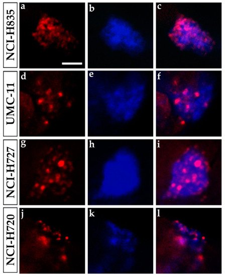
Ki-67 immunostaining of lung carcinoid grafted embryos. Representative images of 48 hpi |
Expression Data
Expression Detail
Antibody Labeling
Phenotype Data
Phenotype Detail
Acknowledgments
This image is the copyrighted work of the attributed author or publisher, and
ZFIN has permission only to display this image to its users.
Additional permissions should be obtained from the applicable author or publisher of the image.
Full text @ Int. J. Mol. Sci.