
Fig. 2
- ID
- ZDB-FIG-220727-27
- Publication
- Chen et al., 2022 - Bridging Size and Charge Effects of Mesoporous Silica Nanoparticles for Crossing the Blood-Brain Barrier
- Other Figures
- All Figure Page
- Back to All Figure Page
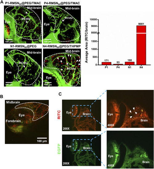
Biodistribution in the brain of four types of RITC-conjugated mesoporous silica nanoparticles (RMSNs) after a pericardial cavity injection in Tg(zfli1:EGFP transgenic zebrafish (200–300 ng/embryo) at 4 days post-fertilization (dpf). (A) Confocal microscopy imaged the distributions of various types of RMSNs in the zebrafish larval brain. The white arrow indicates mesencephalic central arteries (MCtAs). Arrowheads indicate that RMSNs (red) were outside the brain’s blood vessels (green). (B) Two-photon microscopic images and (C) fluorescence microscopic analysis of zebrafish embryo brains after injection with N4-RMSN50@PEG/THPMP. Red (RITC) fluorescence and green (GFP) fluorescence, respectively, represent RMSNs and blood vessels. Arrowheads indicated RMSNs (red). |