
Fig. 3
- ID
- ZDB-FIG-220627-44
- Publication
- Li et al., 2022 - Identification of Novel Vascular Genes Downstream of Islet2 and Nr2f1b Transcription Factors
- Other Figures
- All Figure Page
- Back to All Figure Page
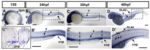
Expression pattern of fsta during zebrafish embryogenesis. (A) The expression of fsta is observed in the lateral plate mesoderm (lpm), the telencephalon (t), diencephalon (d), and hindbrain (h) at the 15S stage embryo. (B,B′) At 24 hpf, fsta is expressed in the head, as well as in vessels (v), and caudal vein plexus (cvp) of the trunk. (B′) is an enlargement of (B). (C,C′) At 30 hpf, fsta expression continues in the head, vessels (v), and CVP of the trunk. (C′) is an enlargement of (C). (C″) Cross sections of embryos from (C′) show that fsta is expressed in the posterior cardinal vein (pcv), and CVP. (D,D′) At 48 hpf, fsta expression in the head, vessels (v), DLAV, and CVP of the trunk. (D′) is an enlargement of (D). Scale bars in all figures represent 200 μm. |