
Fig. 2
- ID
- ZDB-FIG-220608-4
- Publication
- Habicher et al., 2022 - A new transgenic reporter line reveals expression of protocadherin 9 at a cellular level within the zebrafish central nervous system
- Other Figures
- All Figure Page
- Back to All Figure Page
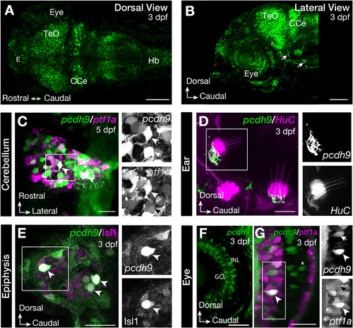
Tg(pcdh9:hs:GFP) expression in the brain of zebrafish larvae. Dorsal (A) and lateral view (B) of the brain of Tg(pcdh9:hs:GFP) zebrafish at 3 days post fertilisation (dpf) show expression in the optic tectum (TeO), cerebellum (CCe), epiphysis (E), in the retina of the eye, the hindbrain (Hb) and axons connecting to the hair cells of the ear (arrow). In the cerebellum pcdh9 was expressed in a subpopulation of Purkinje cells labelled by Tg(ptf1a:dsRed) (arrowhead), ptf1a negative cells are indicated with an arrow (C). In the ear, pcdh9 positive axons connecting to the hair cells, labelled by Tg(HuC:GAL4; UAS:RFP), were observed (D). Immunohistochemistry showed that all pcdh9-eGFP positive cells in the epiphysis were positive for Isl1 (E). The pcdh9-eGFP positive cells in the retina were located in the ganglion cell layer (GCL) and in the inner nuclear layer (INL) (F). In the INL a subset of pcdh9 positive cells were amacrine cells labelled by Tg(ptf1a:dsred) (arrowhead), a subset negative for ptf1a (arrows). Bipolar cells were also pcdh9 positive (G, asterix). Scale bars equal 100 μm in A-B and 20 μm in C-G. |
Reprinted from Gene expression patterns : GEP, 44, Habicher, J., Manuel, R., Pedroni, A., Ferebee, C., Ampatzis, K., Boije, H., A new transgenic reporter line reveals expression of protocadherin 9 at a cellular level within the zebrafish central nervous system, 119246, Copyright (2022) with permission from Elsevier. Full text @ Gene Expr. Patterns