
Fig. 3
- ID
- ZDB-FIG-220608-38
- Publication
- Yoon et al., 2022 - Zebrafish models of Alx-linked frontonasal dysplasia reveal a role for Alx1 and Alx3 in the anterior segment and vasculature of the developing eye
- Other Figures
- All Figure Page
- Back to All Figure Page
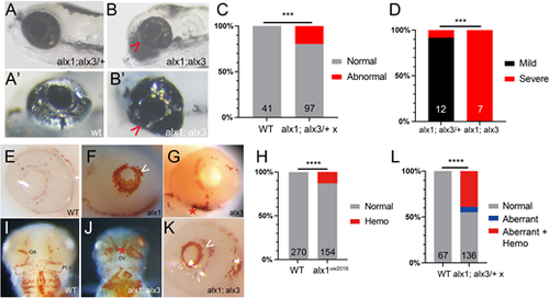
Zebrafish alx1 and alx3 function redundantly during ocular morphogenesis. (A,B) Larvae generated from a cross between alx1;alx3/+ parents were scored at 5 dpf for ocular defects. An alx1;alx3/+ larva with normal ocular morphology (A) similar to that of a wildtype embryo (A′). (B) An alx1;alx3 sibling with ocular coloboma and a misshapen eye (severe defect, arrowheads in B,B′). (C) These ocular defects are restricted to progeny of alx1;alx3/+ parents (***P=0.0009, Fisher's exact test, two trials). (D) The severe ocular defect shown in B is strongly associated with the alx1;alx3 genotype, while the majority of alx1;alx3/+ embryos present with a milder misshapen eye phenotype (not shown) (***P=0.0002 using Fisher's exact test, two trials). (E-L) Embryos derived from wildtype or alx1;alx3/+ parents were stained with o-dianisidine at 2-3 dpf to label hemoglobin in red blood cells. Normal hemoglobin distribution in the eye of a wildtype embryo (E) versus ocular hemorrhage (arrowhead in F) and periocular hemorrhage (asterisk in G) in alx1 mutants. (H) Hemorrhaging is restricted to a subset of alx1 mutants (****P<0.0001, Fisher's exact test, five trials). Normal hemoglobin distribution in the pharyngeal region of a wildtype embryo (I) versus evidence of hemorrhaging in an alx1;alx3 embryo (J). Intraocular and periocular hemorrhages (asterisk) near the optic artery and optic vein in an alx1;alx3 embryo (K). (L) Hemorrhaging is strongly associated with eye defects in alx1;alx3/+ and alx1;alx3 embryos (****P<0.0001, Chi-square test, two trials). Embryos are shown in lateral views, anterior to the left except in I and J, which are ventral views, anterior to the top. OA, optic artery; OV, optic vein; PLV, palatocerebral vein. |
| Fish: | |
|---|---|
| Observed In: | |
| Stage Range: | Protruding-mouth to Day 5 |