
Fig. 1
- ID
- ZDB-FIG-220608-36
- Publication
- Yoon et al., 2022 - Zebrafish models of Alx-linked frontonasal dysplasia reveal a role for Alx1 and Alx3 in the anterior segment and vasculature of the developing eye
- Other Figures
- All Figure Page
- Back to All Figure Page
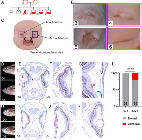
Ocular malformations are associated with ALX1/alx1 loss-of-function in humans and zebrafish. (A) Consanguineous ALX1L165F/+ parents produced 13 children, four of whom were homozygous for ALX1L165F and had complex frontonasal dysplasia (FND3) (subject numbers noted in red). Unaffected individuals did not have eye or facial phenotypes suggestive of FND. (B) Diagram summarizing the anophthalmia, coloboma and bilateral Tessier oblique facial clefts (ObFC) characteristic of FND3. (C) Human subjects displayed bilateral ObFC (not shown) and a range of ocular malformations. The eldest sibling (subject 3) presented with right coloboma and left microophthalmia. The next child (subject 4) presented with bilateral anophthalmia with fused eyelids and shallow orbits. Subject 5 presented with bilateral anophthalmia with open shallow orbits. The upper and lower eyelids were absent, exposing the orbital mucosa. The nasal alae were also malformed with nodular skin tags. Subject 6 had bilateral anophthalmia, fused eyelids, and shallow orbit, similar to subject 4. (D-L) alx1uw2016 adult zebrafish exhibit milder ocular defects, primarily in the anterior ocular segment. (D-G) A representative alx1uw2016 adult with a unilateral ocular malformation (D). (E) Transverse section through the head of the fish in E shows a normal left eye and aphakia (absent lens) on the right. (F) The right eye lacks the lens and the ventral annular ligament but is largely normal otherwise. (G) The right eye contains mononuclear inflammatory cells near the ventral iris and the optic nerve head (asterisks). (H-K) A representative alx1uw2016 zebrafish with bilateral ocular defects (H). (J) In transverse section through the head, the left eye is missing the lens (aphakia) and contains a thick, multifocally disrupted lens capsule, which is embedded in the anterior chamber. There is no annular ligament on the dorsal iridocorneal angle. (K) The right eye contains a small spherical lens nucleus inside a wrinkled lens capsule. The ventral iridocorneal angle is devoid of the annular ligament. (L) Ocular defects were observed in 21.2% of alx1uw2016 homozygotes examined, and none of the wildtype fish of a similar age (****P<0.0001, Fisher's exact test). OD: oculus dexter, right eye; OS: oculus sinister, left eye; al: annular ligament; L: lens; *, mononuclear inflammatory cells. |
| Fish: | |
|---|---|
| Observed In: | |
| Stage: | Adult |