
Fig. 2
- ID
- ZDB-FIG-220607-31
- Publication
- Varela et al., 2022 - Cdkl5 mutant zebrafish shows skeletal and neuronal alterations mimicking human CDKL5 deficiency disorder
- Other Figures
- All Figure Page
- Back to All Figure Page
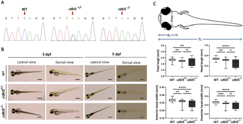
Characterization of cdkl5sa21938 mutant at initial larval stages. (A) Sequencing chromatograms showing the zebrafish genotypes for cdkl5. Arrows indicate the mutation site. (B) Representative lateral and dorsal images of wild-type (WT), heterozygous (cdkl5+/−), and homozygous (cdkl5−/−) mutant embryos with 3 dpf and 5 dpf. Scale bar = 0.5 mm. (C) Morphometric analysis of 5 dpf WT (n = 51), cdkl5+/− (n = 50) and cdkl5−/− (n = 71) embryos. Data are presented as median with interquartile range. Three independent experiments were performed. Statistical analysis was performed using Kruskal–Wallis followed by Dunn multiple comparisons test. *, **, ***, **** indicate p < 0.05, p < 0.01, p < 0.001 and p < 0.0001, respectively. ns indicates not significant. TL-total length; HL-head length; AHW-anterior head width; PHW-posterior head width. |
| Fish: | |
|---|---|
| Observed In: | |
| Stage Range: | Protruding-mouth to Day 5 |