
Fig. 2
- ID
- ZDB-IMAGE-220607-29
- Publication
- Varela et al., 2022 - Cdkl5 mutant zebrafish shows skeletal and neuronal alterations mimicking human CDKL5 deficiency disorder
- All Figures
- Figures for Varela et al., 2022
|
Fig. 2
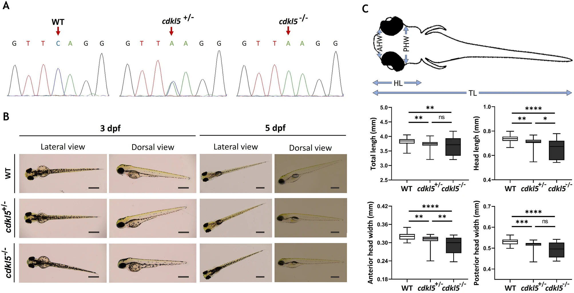
Characterization of cdkl5sa21938 mutant at initial larval stages. (A) Sequencing chromatograms showing the zebrafish genotypes for cdkl5. Arrows indicate the mutation site. (B) Representative lateral and dorsal images of wild-type (WT), heterozygous (cdkl5+/−), and homozygous (cdkl5−/−) mutant embryos with 3 dpf and 5 dpf. Scale bar = 0.5 mm. (C) Morphometric analysis of 5 dpf WT (n = 51), cdkl5+/− (n = 50) and cdkl5−/− (n = 71) embryos. Data are presented as median with interquartile range. Three independent experiments were performed. Statistical analysis was performed using Kruskal–Wallis followed by Dunn multiple comparisons test. *, **, ***, **** indicate p < 0.05, p < 0.01, p < 0.001 and p < 0.0001, respectively. ns indicates not significant. TL-total length; HL-head length; AHW-anterior head width; PHW-posterior head width.