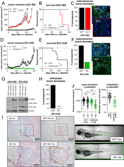
FGFR4 impacts on tumorigenicity and tumor growth. A-F SCID/CB17 mice were subcutaneously injected with wild-type FGFR4-388Gly-expressing, endogenously FGFR4low U251-MG cells or GFP-transduced controls (n = 4/group) (A-C) or kinase-dead FGFR4-KD(K504M)-expressing, endogenously FGFR4high BTL1528 cells or GFP-transduced controls (n = 7/group) D-F. Tumor growth curves (mean tumor volume ± SEM). + = mouse sacrificed (A/D), Kaplan Meier survival curves (B/E), tumor take (C/F, left), and representative cryo-sections of DAPI stained tumor xenografts (C/F, right) are shown. G Western blot analysis of the indicated proteins in U251-MG (left) or BTL1528 (right) FGFR4-altered xenografts (compare panels A-F). β-actin served as loading control. H Orthotopic tumor formation of BTL1528 GFP and FGFR4-modulated sublines in mice (n = 5/group) is shown. (I) Representative photomicrographs of a BTL1528 GFP orthotopic tumor stained with HE (left panels) or GFP by IHC (right panels). J Primary tumor area over time (left) and extra-primary tumor cell migration in zebrafish larvae (right) of BTL1528 GFP- or FGFR4-KD(K504M)-expressing models one-day post injection (dpi). Data from the independent experiments are shown. Representative photomicrographs of zebrafish larvae are depicted in the lower panels. black arrows: primary tumors, white arrows: extra-primary site tumor cell clusters; 2-way ANOVA (A + D; J left), log-rank (Mantel-Cox) test (B + E), or Student’s t-test (J right). n.d. = no detectable tumors, n.s. = not significant, *p < 0.05, ***p < 0.001. GLY = FGFR4-388Gly, KD = FGFR4-KD(K504M), s.c. = subcutaneous, short/long: exposure times
|
