|
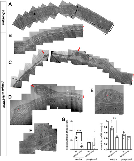
Electron microscopy analysis of cornea. Electron microscopy data for 14-dpf wild-type, A, and mab21l1c.107delA mutant, B-F, corneas. Please note thinner (B,C,D; red brackets) and unevenly folded (D; red arrowhead) corneas, especially epithelial layer, in mutants vs wild-type, as well as the high number of abnormal cells (cells undergoing cell death [red arrows in, C], goblet [red asterisks in, C, F], and possible rodlet cells [red asterisks in D, E inset]); bar = 2 μM; (ce) corneal epithelium; (cs) corneal stroma; (cn) corneal endothelium; (le) lens epithelium. G, Measurements of corneal epithelial and stromal thickness for central and peripheral regions comparing wild-type and mab21l1c.107delA fish at 14-dpf based on EM images. Statistical significance is indicated by asterisks (central epithelium ***P = .00012; central stroma **P = .0018); error bars indicate SEM
|