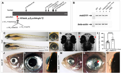
MAB21L1/mab21l1 variants and gross phenotypic analysis of mab21l1 deficient fish. A, Schematic of known human MAB21L1 pathogenic variants (top) and zebrafish mab21l1 alleles generated in this study (bottom). The red arrow indicates the zebrafish line chosen for detailed phenotypic characterization, mab21l1-c.107delA p.(Lys36Argfs*7). B, RT-PCR analysis of mab21l1 transcript expression comparing 5-dpf wild-type (lanes 1 and 2) and homozygous mab21l1-c.107delA p.(Lys36Argfs*7) (lanes 3 and 4) embryos. Beta-Actin was used as a loading control. C-I, Assessment of 5-dpf phenotypes. No gross abnormalities between wild-type, C,E, and mab21l1c.107delA embryos, D,F, were detected in lateral position. However, an increase in anterior chamber area in mab21l1c.107delA embryos (G, H, indicated by red arrowheads in, H) was detected in dorsal view. This finding was quantified, I, and found to be statistically significant (****P = .0001); error bars indicated SEM. J-O, Assessment of adult phenotypes (1–2 years of age). Smaller eyes with small/misshapen pupils, M, N, corneal opacities (N; red arrowhead), and irregularly shaped lens with keratolenticular adhesions, O, were observed. (c) cornea, (l) lens, (r) retina
|
