|
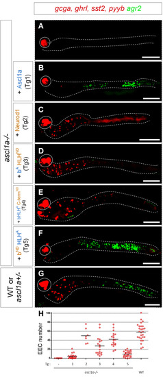
Analysis of rescuing capacities of chimeric Neurod1/ Ascl1a proteins in <italic toggle='yes'>ascl1a-/-</italic> larvae.A-G: FISH performed at 96 hpf with the agr2 probe revealed in green and a mix of hormones probes (ghrl, pyyb, gcga and sst2) revealed in red on ascl1a -/- larvae without (A) or with Tg1 to Tg5 transgenes (B to F) and on sibling control embryos (G). All the larvae have been heat-shocked at 36, 46 and 56 hpf. The transgenic line used is indicated on the left part of the figure as well as the genotype of the larvae; the ascl1a-/- larvae were identified by the loss of the pituitary prl expression (not shown). All views are ventral with the anterior part to the left and represent confocal projection images. The pancreas is encircled while the location of the gut, visualised with a DAPI staining (not shown), is delimited by dashed lines. Scale bar: 100μm. H: Quantification of the number of ghrl+/pyyb+/gcga+/sst2+ EEC detected in conditions A to G counted under a fluorescent microscope.
|