
FIGURE
Figure 4
- ID
- ZDB-FIG-220329-18
- Publication
- Alkowari et al., 2022 - Functional Characterization of the MYO6 Variant p.E60Q in Non-Syndromic Hearing Loss Patients
- Other Figures
- All Figure Page
- Back to All Figure Page
Figure 4
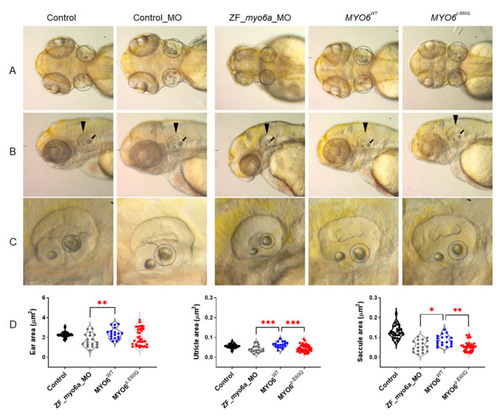
Figure 4. Detection of ear morphological defects in zebrafish models. (A) Dorsal view of zebrafish at 72 hpf. (B) Lateral view of the posterior otolith at 72 hpf zebrafish (black arrow). (C) Closeup view of zebrafish ear morphology (black dashed circle). (D) The perimeter length of zebrafish ear, utricle, and saccule area in μm2. The total number of experiments was 3, and the number of embryos analyzed was 31, 18, 19, and 36 for control, ZF_myo6a_MO, MYO6WT, and MYO6p.E60Q groups, respectively. One-way ANOVA using GraphPad Prism software (version 8.0) and Tukey’s multiple comparisons test with p-values of * p < 0.05, ** p < 0.01, *** p < 0.001.
|
Expression Data
Expression Detail
Antibody Labeling
Phenotype Data
| Fish: | |
|---|---|
| Knockdown Reagent: | |
| Observed In: | |
| Stage: | Protruding-mouth |
Phenotype Detail
Acknowledgments
This image is the copyrighted work of the attributed author or publisher, and
ZFIN has permission only to display this image to its users.
Additional permissions should be obtained from the applicable author or publisher of the image.
Full text @ Int. J. Mol. Sci.