
Fig. 4
- ID
- ZDB-FIG-220304-35
- Publication
- Wyatt et al., 2021 - Post-translational activation of Mmp2 correlates with patterns of active collagen degradation during the development of the zebrafish tail
- Other Figures
- All Figure Page
- Back to All Figure Page
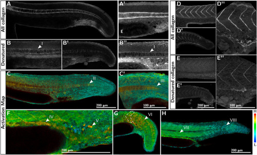
Activated Mmp2 co-localizes with denatured collagen in early development. CHP-mediated detection of total collagen (A, A′) consistently reveals abundant collagen in the notochord sheath and notochord cells (n = 10). The collagen in the notochord sheath at the yolk extension is 1–2 μm thick and fibrous (A′). Endogenously denatured collagen is found in the notochord (B), especially in the sheath (I), but also in between the notochord cells (B′). Activation of EMMA-Mmp2 in the notochord (C) was visible in isolated 'hot spots' along the trunk in 18 of 96 embryos imaged (II), that were in register with myotome boundaries in 9 of 25 instances. Activation of EMMA-Mmp2 is consistently observed in the notochord sheath (III), especially in the posterior tail (C′). Collagen is always present in somite boundaries almost as soon as they are distinguishable (D), and includes a small amount of denatured collagen at this stage (E). Activation of EMMA-Mmp2 is usually (74%) associated with the gut epithelium (IV) and we observed activation in ventral caudal hematopoietic tissue (V) in 80% of embryos. In all embryos examined (n = 27) we see activation in somite boundaries (VI), which is always stronger in anterior somite boundaries (VII) compared with posterior somites (VIII). |
Reprinted from Developmental Biology, 477, Wyatt, R.A., Crawford, B.D., Post-translational activation of Mmp2 correlates with patterns of active collagen degradation during the development of the zebrafish tail, 155-163, Copyright (2021) with permission from Elsevier. Full text @ Dev. Biol.