|
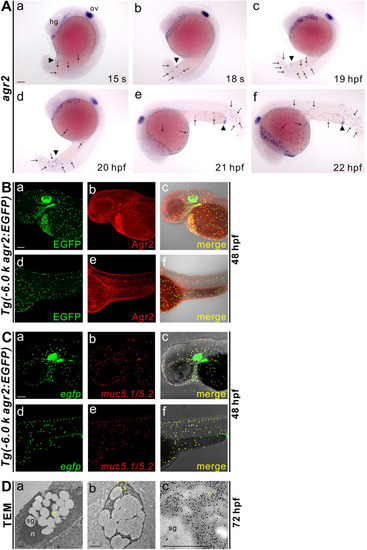
Characterization of agr2+ epidermal mucous cells. (A). Developmental expression pattern of agr2 in the epidermis from 15 s to 22 hpf. hg, hatching gland; ov, otic vesicle. Arrows, EMCs. Arrowhead, anus. Scale bar, 100 μm. (B). Colocalization of Agr2 (red) and EGFP (green) was detected in the trunk and yolk regions of Tg(-6.0 k agr2:EGFP) embryos at 48 hpf. Scale bar, 100 μm. (C). egfp (green) is coexpressed with muc5.1/muc5.2 (red) in Tg(-6.0 k agr2:EGFP) embryos at 48 hpf. Scale bar, 100 μm. (D). Ultrastructure of EMCs and GFP immuno labeling in Tg(-6.0 k agr2:EGFP) embryos at 72 hpf. n, nucleus; sg, secretory granules. Yellow arrowhead, endoplasmic reticulum. Yellow square box indicates enlarged area. Scale bars, 1 μm. Scale bar for enlarged image (c), 0.5 μm.
|